 Advanced equipment
The core provides advanced transmitted-light and fluorescence microscopy equipment paired with flow cytometry and cell sorting equipment.
Equipment
Flow cytometers
- BD FACSCanto X clinical flow cytometry system. This system provides a four-laser analytical platform with a 40-tube loader. It uses colinear 4,050-nanometer and 640-nanometer lasers together with 488-nanometer and 561-nanometer excitation to support comprehensive multicolor detection. The system captures up to 13 fluorescence parameters in addition to forward and side angle light scatter, with customizable filters to accommodate a wide range of emission wavelengths. A 40-tube automated sample loader enables walk-away operation, increasing throughput while reducing hands-on time. Designed for efficient workflow integration, the system combines reliable fluidics and optics with intuitive software control to streamline setup, acquisition and data management for both routine and advanced flow cytometry applications.
- BD FACSDiscover S8. This real-time imaging-based sorting and spectral analyzer provides precise, visually confirmed cell sorts. It elevates research by being the first system to combine high-parameter spectral flow cytometry with breakthrough BD CellView Image Technology, allowing users to sort what they see in real time. This revolutionary imaging-based sorting capability empowers users to isolate cells with far greater precision than traditional methods by using spatial and morphological insights, such as protein localization, cell shape or viral distribution, to identify and sort target populations that were previously indistinguishable. It is equipped with four lasers and 48 fluorescent detectors plus three imaging detectors. The BD FACSDiscover S8 provides the ultimate in panel flexibility and data clarity, ensuring that every sorted population is visually confirmed for higher confidence in downstream applications.
- BD FACSMelody cell sorter. This user-friendly three-laser cell sorter features automated setup and rapid single cell deposition into plates. It is designed for efficiency and convenience, featuring an automated setup process that significantly reduces preparation time. The system is ready for sorting in less than 20 minutes. The cell sorter is operated with intuitive, wizard-driven BD FACSChorus Software. It is easy to learn and straightforward to use. This three-laser sorter detects up to eight fluorescence parameters and uses a 100-micron nozzle, supporting simultaneous sorting of two or four populations. It enables precise single-cell deposition into 96-well plates for cloning, 384-well polymerase chain reaction (PCR) plates or other compatible collection devices. Users can be trained to independently operate the FACSMelody cell sorter, allowing for flexible access to sorting services. This includes after-hours and weekend use.
- BD LSRFortessa X-20 Cell Analyzer. This high-performance five-laser cell analyzer is optimized for 20-parameter experiments. It is a high-performance analytical (nonsorting) digital flow cytometer designed for fast, efficient and highly sensitive multicolor analysis. This customized five-laser system detects up to 20 fluorescence parameters and is equipped with state-of-the-art lasers and advanced optics to deliver excellent signal resolution for complex multicolor experiments. This cell analyzer features a fluidics wet cart that enhances fluidic stability and allows fluids to be changed during operation without interrupting data acquisition, supporting reliable performance and efficient, uninterrupted workflows for both routine and advanced flow cytometry applications.
- Bio-Rad ZE5 Cell Analyzer. This five-laser cell analyzer features a universal plate loader, temperature control and a sample-return function. It is a next-generation analytical digital flow cytometer designed for fast, high-efficiency data acquisition with integrated temperature control. It detects up to 30 fluorescence parameters in addition to forward and side scatter, delivering high sensitivity through state-of-the-art optics and laser technology. The cell analyzer features a universal plate loader compatible with single tubes, full racks of 5-milliliter or 1.5-milliliter tubes, and 96- or 384-well plates, enabling flexible and efficient sample handling. A built-in adjustable orbital vortex shaker provides effective mixing and active temperature regulation, while allowing unused samples to be returned to the original tube or well. Onboard fluidics enhance system stability and support fluid changes during operation without interrupting data acquisition. This ensures efficient and uninterrupted workflows.
- Cytek Aurora spectral analyzer. This four-laser full spectral cytometer provides high-resolution, 48-parameter analysis and autofluorescence extraction. It uses a sophisticated four-laser array to detect up to 48 parameters with unmatched precision. By capturing the complete emission spectrum of every molecule and offering advanced autofluorescence extraction, this system provides superior resolution for small particles and high-dimensional panels, all while supporting high-throughput workflows via a versatile plate loader and integrated orbital vortex shaker.
- ZetaView nanoparticle tracker. This dual-laser nanoparticle tracker simultaneously measures size, concentration and zeta potential in exosomes and viruses. The ZetaView TWIN Nanoparticle Tracking Analyzer features dual 488-nanometer and 640-nanometer lasers and provides comprehensive, individual-particle characterization by simultaneously measuring hydrodynamic size, concentration and zeta potential. It is optimized for biological nanoparticles such as extracellular vesicles, exosome and viruses. This high-resolution system uses automated scanning across 11 focal positions to ensure superior statistical accuracy. The system enables advanced fluorescence detection to precisely identify and colocalize labeled subpopulations within complex samples.
All cell sorters can sort up to four populations simultaneously. Single cells can be sorted into each well of a 96-well plate for cloning, into 384-well PCR plates or into other collection devices. Most analytical flow cytometers are equipped with an automatic sample loader or plate loader for convenient sample handling. The ZE5 and FX-20 models also feature a temperature control system that maintains sample temperatures between 4 degrees Celsius and 40 degrees Celsius.
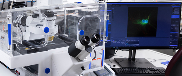
Optical imaging equipment
- Alpenglow 3Di. This top-load, hybrid open-top light-sheet system is designed for 3D imaging of large specimens or multiwell plate samples, enabling volumetric visualization and analysis.
- Axioplan 2 with Jenoptik camera. This provides standard digital brightfield imaging.
- Confocal laser scanning microscopes. These microscopes use a focused laser beam to scan samples point by point. The ability of confocal microscopy to eliminate out-of-focus light by employing a pinhole aperture results in excellent optical sections with high contrast and enables the high-resolution reconstruction of 3D structures. In the life sciences, confocal microscopes are used to visualize structures within cells and tissues, study dynamic biological processes, and analyze multifluorescent samples.
- LSM Lightfield 4D — instant volumetric imaging. The system can capture complete volumetric datasets in a single acquisition, enabling real-time observation of dynamic biological processes. This capability is ideal for live-cell and whole-organism imaging where speed and gentle illumination are critical.
- LSM Spectral Multiplex — full-range spectral imaging. The LSM Spectral Multiplex module supports precise spectral separation across the full fluorescence wavelength range, making it possible to distinguish numerous fluorophores — even those with overlapping spectra. This enables advanced multiplex labeling experiments with clean, accurate signal separation.
- Oxford Instruments BC-43 widefield and confocal benchtop microscope. The BC43 is a compact, high-performance benchtop imaging system that brings advanced widefield, confocal and super-resolution microscopy into an easy-to-use, space-saving design. It delivers high-quality 2D and 3D imaging across a broad range of samples from thin cultured cells to thick tissues and organoids. It includes transmitted light modes (brightfield and differential phase contrast) for label-free contrast. The system integrates Fusion Benchtop software for streamlined multimodal acquisition and Imaris software for powerful 3D visualization and analysis.
- BC43 confocal. The BC43 confocal incorporates technology from the award-winning Dragonfly platform to deliver fast, flexible and high-quality 3D imaging. Users can image deep into thick samples, capture rapid live events with minimal phototoxicity and acquire large multiposition datasets using the motorized XYZ stage. Confocal imaging is more than 10 times faster than traditional point-scanning systems, enabling long-term live imaging with outstanding clarity.
- BC43 widefield. The BC43 widefield is ideal for light-sensitive samples and high-speed imaging of dynamic processes. It offers exceptional speed, high sensitivity for low-signal specimens and improved resolution through Imaris ClearView-GPU deconvolution.
- Super-resolution with SRRF-Stream+. SRRF-Stream+ provides fast, one-click super-resolution compatible with both widefield and confocal imaging. It supports standard labeling protocols, a wide range of sample types and live imaging workflows, achieving resolutions 1.5 times to 1.7 times beyond the diffraction limit down to approximately 140 nanometers.
- Transmitted light imaging. Brightfield and differential phase contrast provide high-contrast, label-free imaging without phototoxicity. These modes reveal fine structural detail and full organism boundaries. They can be easily overlaid with fluorescence data for richer spatial context.
- Zeiss Lightsheet 7. This light-sheet fluorescence microscopy solution is used for rapid, gentle imaging of whole organisms, tissues and long-term live samples. It is a versatile light-sheet fluorescence microscope designed for fast, gentle 3D imaging of living samples and large cleared tissues. Its dedicated optics, sample chamber and holders adapt to a wide range of refractive indices and clearing methods, enabling whole-specimen imaging — up to 2 centimeters in size — with subcellular resolution. High-efficiency detectors allow rapid imaging at extremely low illumination levels, preserving sample health during long-term live experiments. For cleared tissues, Lightsheet 7 supports nearly all common clearing solutions and provides precise environmental control for live imaging. Advanced features such as adaptive optics, flexible chamber configurations and Zeiss' patented Pivot Scan technology ensure artifact-free optical sections and outstanding image quality. Smart software tools streamline setup, tiling, multiview imaging and data processing, making the Lightsheet 7 ideal for organoids, spheroids, whole organs, developing organisms and other large 3D specimens.
- Zeiss LSM 780. This solution offers confocal imaging with Z-stack, tile scan and time-lapse capability. Environmental control is available.
- Zeiss LSM 980 Airyscan 2. This confocal and super-resolution imaging solution with multiplex capability supports Z-stacks, tile scans, time-lapse imaging and environmental control (temperature, carbon dioxide and humidity). This high-sensitivity detector system for confocal and super-resolution imaging is designed to provide fast acquisition, improved signal-to-noise and enhanced spatial resolution. Its detector array enables gentle, high-speed imaging while resolving fine subcellular structures and supporting multiplexed fluorescence experiments across a wide range of sample types — from single cells to whole living organisms.
Scanners
- KFBIO MagScanner KF-PRO-005. This digital pathology slide scanner can image up to five slides a run. It supports both brightfield and fluorescence modalities. The system provides automated slide loading and high-resolution whole-slide imaging.
- Zeiss Axio Scan.Z1. This fully automated high-throughput digital slide scanner can image up to 100 slides a run. It supports both brightfield and fluorescence modalities. It's designed for reliable, hands-off imaging of large sample sets. The scanner is built for modern research workflows that demand speed, consistency and scalability. It offers flexible configuration options to accommodate diverse sample types and evolving experimental needs. With automated slide loading, 24/7 operation and proven Zeiss optical components, the Axio Scan.Z1 delivers high-quality virtual slides, including fast, high-resolution fluorescence imaging, with minimal user intervention. It streamlines acquisition, analysis and data management, making it ideal for both routine tasks and complex, data-intensive microscopy applications.