 Broad range of microscopy platforms
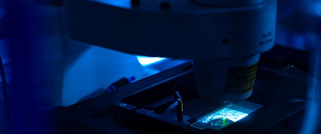
The core provides a broad range of transmitted-light and fluorescence microscopy platforms, including widefield, confocal, super-resolution and light-sheet imaging.
Optical microscopy imaging equipment
The core provides a broad range of transmitted-light and fluorescence microscopy platforms, including widefield, confocal, super-resolution and light-sheet imaging. Off-peak hour use is available at reduced rates. For more information, email the core.
Imaging equipment includes:
Alpenglow 3Di
This top-load, hybrid open-top light-sheet system is designed for 3D imaging of large specimens or multiwell plate samples. This system enables volumetric visualization and analysis.
Axioplan 2 with Jenoptik camera
This is used for standard digital brightfield imaging.
Oxford Instruments
- BC43. The BC43 is a compact, high-performance benchtop imaging system that brings advanced widefield, confocal and super-resolution microscopy into an easy-to-use, space-saving design. It delivers high-quality 2D and 3D imaging across a broad range of samples from thin cultured cells to thick tissues and organoids. It includes transmitted light modes (brightfield and differential phase contrast) for label-free contrast. The system integrates Fusion Benchtop software for streamlined multimodal acquisition and Imaris software for powerful 3D visualization and analysis.
- BC43 confocal. The BC43 confocal incorporates technology from the award-winning Dragonfly platform to deliver fast, flexible and high-quality 3D imaging. Users can image deep into thick samples, capture rapid live events with minimal phototoxicity and acquire large multiposition datasets using the motorized XYZ stage. Confocal imaging is more than 10 times faster than traditional point-scanning systems, enabling long-term live imaging with outstanding clarity.
- BC43 widefield. The BC43 widefield is ideal for light-sensitive samples and high-speed imaging of dynamic processes. It offers exceptional speed, high sensitivity for low-signal specimens and improved resolution through Imaris ClearView-GPU deconvolution.
- Super-resolution with SRRF-Stream+. SRRF-Stream+ provides fast, one-click super-resolution compatible with both widefield and confocal imaging. It supports standard labeling protocols, a wide range of sample types and live imaging workflows, achieving resolutions 1.5 times to 1.7 times beyond the diffraction limit down to approximately140 nanometers.
- Transmitted light imaging. Brightfield and differential phase contrast provide high-contrast, label-free imaging without phototoxicity. These modes reveal fine structural detail and full organism boundaries. They can be easily overlaid with fluorescence data for richer spatial context.
Zeiss Lightsheet 7
Light-sheet fluorescence microscopy is used for rapid, gentle imaging of whole organisms, tissues and long-term live samples.
The Zeiss Lightsheet 7 is a versatile light-sheet fluorescence microscope designed for fast, gentle 3D imaging of living samples and large cleared tissues. Its dedicated optics, sample chambers and holders adapt to a wide range of refractive indices and clearing methods, enabling whole-specimen imaging — up to 2 centimeters in size — with subcellular resolution.
High-efficiency detectors allow rapid imaging at extremely low illumination levels, preserving sample health during long-term live experiments. For cleared tissues, Lightsheet 7 supports nearly all common clearing solutions and provides precise environmental control for live imaging.
Advanced features such as adaptive optics, flexible chamber configurations and Zeiss' patented Pivot Scan technology ensure artifact-free optical sections and outstanding image quality. Smart software tools streamline setup, tiling, multiview imaging and data processing, making the Lightsheet 7 ideal for organoids, spheroids, whole organs, developing organisms and other large 3D specimens.
Zeiss LSM 780
This solution offers confocal imaging with Z-stack, tile scan and time-lapse capability. Environmental control is available.
Confocal laser scanning microscopes use a focused laser beam to scan samples point by point. The ability of confocal microscopy to eliminate out-of-focus light by employing a pinhole aperture results in excellent optical sections with high contrast and enables the high-resolution reconstruction of 3D structures. In life sciences, confocal microscopes are used to visualize structures within cells and tissues, study dynamic biological processes, and analyze multifluorescent samples.
Zeiss LSM 980 Airyscan 2
This confocal and super-resolution imaging solution provides multiplex capability. It supports Z-stacks, tile scans, time-lapse imaging and environmental control (temperature, carbon dioxide and humidity).
This microscope has these capabilities:
- Airyscan 2. This high-sensitivity detector system for confocal and super-resolution imaging is designed to provide fast acquisition, improved signal-to-noise and enhanced spatial resolution. Its detector array enables gentle, high-speed imaging while resolving fine subcellular structures and supporting multiplexed fluorescence experiments across a wide range of sample types from single cells to whole living organisms.
- LSM Lightfield 4D — instant volumetric imaging. The system can capture complete volumetric datasets in a single acquisition, enabling real-time observation of dynamic biological processes. This capability is ideal for live-cell and whole-organism imaging where speed and gentle illumination are critical.
- LSM Spectral Multiplex — full-range spectral imaging. The LSM Spectral Multiplex module supports precise spectral separation across the full fluorescence wavelength range, making it possible to distinguish numerous fluorophores — even those with overlapping spectra. This enables advanced multiplex labeling experiments with clean, accurate signal separation.
Consultations
Email the core.