 Disease-associated extracellular vesicles isolated from human brain tissue
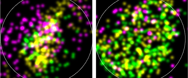
This direct stochastic optical reconstruction microscopy image shows punctate staining of phosphorylated tau (cyan), total tau (yellow) and pan-tetraspanin (magenta) loaded on a single brain-derived extracellular vesicle from patients with Alzheimer's disease. At left is the apolipoprotein E (APOE) 3/3 genotype. At right is the APOE 4/4 genotype.
Tau propagation mechanisms
Dr. Ikezu has been a pioneer in defining the roles of microglia and extracellular vesicles in the spread of pathogenic tau protein in the brain. His group elegantly demonstrated how microglia phagocytose tau-containing neurons secrete extracellular vesicles containing tau protein to accelerate tau pathology using the adeno-associated virus-mediated gene transfer system. In 2016, the publication of this discovery was recognized by the Alzheimer's Association as the most impactful paper of the previous two years.
Dr. Ikezu's laboratory also showed that brain-derived extracellular vesicles propagate tau pathology compared with oligomer or fibrillar tau aggregates. The lab discovered that Wolframin-1+ pyramidal neurons in the layer II entorhinal cortex are responsible for the propagation of tau from the brain region to the CA1 region of the hippocampal field. This is commonly seen in primary age-related tauopathy and the early stage of a brain affected by Alzheimer's disease.
References
- Delpech JC, Pathak D, Varghese M, Kalavai SV, Hays EC, Hof PR, Johnson WE, Ikezu S, Medalla M, Luebke JI, Ikezu T. Wolframin-1-expressing neurons in the entorhinal cortex propagate tau to CA1 neurons and impair hippocampal memory in mice. Science Translational Medicine. 2021; doi:10.1126/scitranslmed.abe8455.
- Ruan Z, Pathak D, Venkatesan Kalavai S, Yoshii-Kitahara A, Muraoka S, Bhatt N, Takamatsu-Yukawa K, Hu J, Wang Y, Hersh S, Ericsson M, Gorantla S, Gendelman HE, Kayed R, Ikezu S, Luebke JI, Ikezu T. Alzheimer's disease brain-derived extracellular vesicles spread tau pathology in interneurons. Brain. 2021; doi:10.1093/brain/awaa452.
- Asai H, Ikezu S, Tsunoda S, Medella M, Luebke J, Haydar T, Wolozin B, Butovsky O, Kügler S, Ikezu T. Depletion of microglia and inhibition of exosome synthesis halt tau propagation. Nature Neuroscience. 2015; doi.org/10.1038/nn.4132.
Learn more
Learn more about our work.