Mitochondrial Dynamics In Health and Disease
Dividing mitochondria in HeLa cells
Dividing mitochondria in HeLa cells
Dividing mitochondria in HeLa cells
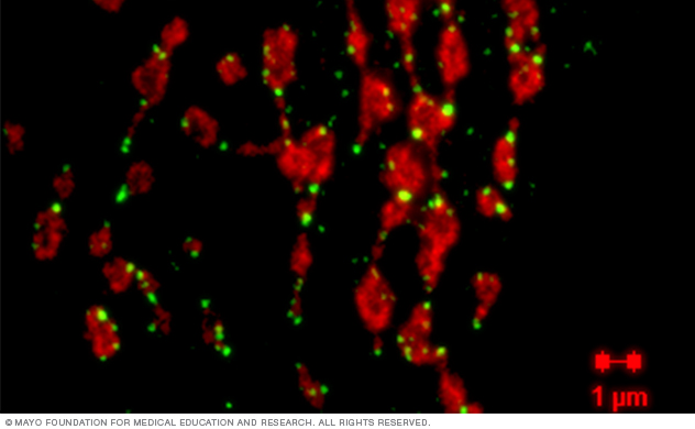
In this image, red shows mitochondria and green shows fission protein Drp1. Visualization of mitochondrial fission proteins allows researchers to determine how dynamic changes in mitochondrial fission and fusion in response to stress or environmental toxicants contribute to health and disease.
In this project, our lab is studying how mitochondria react to stress associated with the development of neurodegenerative diseases and aging.
Assessment of mitochondrial motility along axons and dendrites and evaluation of mitochondrial fission and fusion in vitro and in vivo helps scientists understand early molecular mechanisms of various diseases and conditions. Better understanding can help advance new strategies for early intervention. Diseases and conditions that could benefit include Alzheimer's disease, Parkinson's disease, Huntington's disease, multiple sclerosis, metabolic disorders and aging.
One example of a combinatorial therapeutic approach is applying a compound that blocks stress-induced mitochondrial fission and subsequent removal via selective mitochondrial degradation (mitophagy). At the same time, another compound could be used to promote mitochondrial biogenesis to replace damaged organelles.
Axonal trafficking of mitochondria in primary neuron from a wild-type mouse
This video offers a visualization of mitochondria in E17 hippocampal neurons. Axonal trafficking of mitochondria is impaired in neurodegenerative diseases and aging. Restoration of axonal trafficking promotes mitochondria distribution within the axons delivering energy to the distal parts, which supports synaptic activity and could preserve memory function.
Our team conducted visualization using a mitochondria-specific dye called tetramethylrhodamine methyl ester (TMRM). We acquired 600 frames by imaging the axon every second using an LSM 510 confocal microscope. Imaging focused on the axon, with the cell body located at the top of the image. The resulting video was analyzed using Analyze, a comprehensive multidimensional medical image processing, visualization and analysis software package developed by the Biomedical Imaging Resource Core.
By treating the microscope image sequence as a spatial stack of cross-sectional images, the volume-rendering algorithms in Analyze produced a 3D digital kymograph. This allowed the motion of multiple organelles over a period of time to be visualized in a single, static 2D image. This final kymograph enables our lab to trace each mitochondrion through all 600 frames to generate a final profile of movement.