 Renal oxidative stress increases with polycystic kidney disease progression
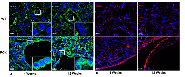
Shown is representative immunofluorescence staining for 8-hydroxyguanosine (green) and NOX4 (red) in renal tissue sections from wild-type and PCK rats at 4 and 12 weeks of age. PCK kidneys demonstrated increased 8-hydroxyguanosine (A) and NOX4 (B) immunoreactivity, compared with wild-type kidneys, with further increases observed as disease progressed.
Oxidative stress and redox signaling
Oxidative stress and abnormal redox signaling are increasingly recognized as important contributors to renal injury in autosomal dominant polycystic kidney disease. The lab investigates how reactive oxygen species, NOX4 signaling, mitochondrial dysfunction and antioxidant responses influence cyst growth, vascular injury and disease progression.
Studies aim to identify mechanisms underlying oxidative stress early in disease and evaluate potential therapeutic strategies targeting redox pathways.
Research areas include:
- NOX4 signaling in autosomal dominant polycystic kidney disease.
- Redox regulation and oxidative injury.
- Antioxidant response pathway.
- Association between reactive oxygen species and vascular dysfunction.
- Biomarkers of oxidative stress in patients with autosomal dominant polycystic kidney disease.
Featured publications
- Irazabal MV, Torres VE. "Reactive Oxygen Species and Redox Signaling in Chronic Kidney Disease." Cells. 2020.
- Kahveci AS, Barnatan TT, Kahveci A, Adrian AE, Arroyo J, Eirin A, Harris PC, Lerman A, Lerman LO, Torres VE, Irazabal MV. "Oxidative Stress and Mitochondrial Abnormalities Contribute to Decreased Endothelial Nitric Oxide Synthase Expression and Renal Disease Progression in Early Experimental Polycystic Kidney Disease." International Journal of Molecular Sciences. 2020.