Real-time in vivo imaging
Dr. Singh's Pancreatitis and Acute Outcomes Research Lab uses novel metabolic tracers in real time to study metabolic demand during pancreatitis. These include near-infrared imaging of 2-deoxyglucose. Whole-body scans show pancreatic uptake in vivo and ex vivo pancreatic uptake. These images show that increased uptake implies "glucose hunger," foreshadowing the severe necrosis that develops later during glyceryl trilinoleate-induced pancreatitis.
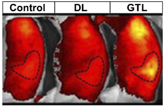 Pancreatic near-infrared 2-deoxyglucose uptake
This image shows pancreatic near-infrared 2-deoxyglucose uptake in pancreatic duct legation-induced mild pancreatitis and severe pancreatitis due to glyceryl trilinoleate infusion.
We can identify the cellular location of such tracers histologically. This allows us to identify the cells with an increase in metabolic demand during severe pancreatitis. Similar approaches can be used to trace lipid behavior, organ failure and sterile to septic transition. Additional methods, such as echocardiography, are used to study cardiac function during the progression of shock in severe pancreatitis.
Videos of an echocardiogram during severe pancreatitis (left) compared with a normal echocardiogram (right) from the Pancreatitis and Acute Outcomes Research Lab.
With these studies, we can investigate the pathophysiology of pancreatitis and acute diseases in a clinically relevant manner.