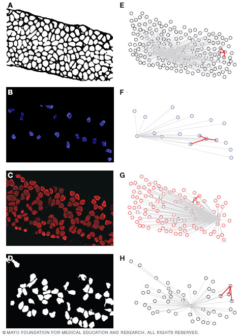
 Diaphragm muscle fibers identified according to fiber type (based on myosin heavy chain isoform expression)
This image shows representative diaphragm muscle cross-sections with type I fibers in blue, type IIa fibers in purple, and type IIx or type IIb fibers both in black. Scale bar is 50 μm. Individual muscle fibers are automatically thresholded and separated for analysis. Greising SM, et al. Analysis of muscle fiber clustering in the diaphragm muscle of sarcopenic mice. Muscle & Nerve. 2015;52:76.
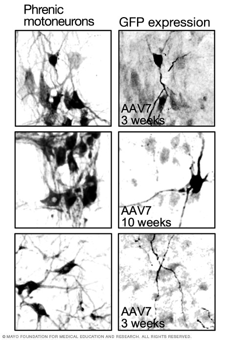 Gene transfer to respiratory motoneurons
This image shows how adeno-associated virus (AAV) efficiently and selectively transduces phrenic motoneurons after intrapleural injection. The left panels show retrogradely labeled motoneurons. The right panels show green fluorescent protein (GFP) expression in both cell bodies (top panels; at 3 and 10 weeks) and dendrites (lower panel).