Research projects
Research in the Developmental Endoscopy Laboratory is focused on creating innovative devices and techniques in endoscopic ultrasound to diagnose and treat a broad range of gastrointestinal conditions, including cancer and obesity.
Our lab has several ongoing initiatives, including four main research projects.
Interventional endoscopic ultrasound
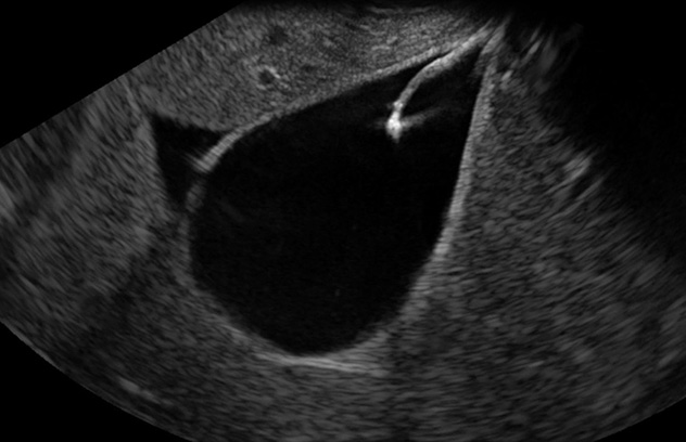
This image shows a fixation device being deployed into a porcine gallbladder under endoscopic ultrasound guidance.
The field of interventional endoscopic ultrasound is rapidly evolving with the advent of novel procedures to treat conditions that previously required surgery.
Our lab is developing techniques and devices to make these procedures safer and more effective. One example of this is the use of an endoscopic ultrasound (EUS)-guided suture device for luminal apposition of the gallbladder to facilitate EUS-guided gallbladder drainage.
Key references
Transmural gallbladder drainage using a novel endosonographic-guided suture (with video). Gastrointestinal Endoscopy. 2025.
Optimizing tissue acquisition with endoscopic ultrasound
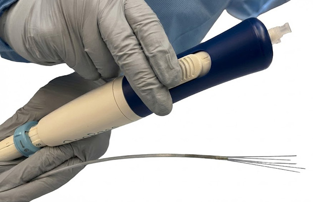
This image shows a diagram of a novel, multipronged needle to facilitate tissue acquisition and treatment during endoscopic ultrasound.
Endoscopic ultrasound is the gold standard to diagnose pancreatic cancer. Sampling with current devices can result in inadequate tissue to obtain an accurate diagnosis, which can lead to repeated procedures and delays in treatment.
Preclinical testing was performed on a novel multipronged needle that may be used to optimize diagnostic sampling and deliver treatments directly to the pancreas in the future. Device optimization and preclinical testing were followed by Food and Drug Administration (FDA) approval. First-in-human studies are now planned.
Key references
Irreversible electroporation and endoscopic treatment of colorectal cancer
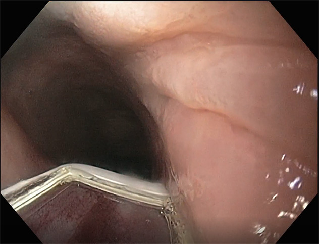
This image shows mucosal ablation of an esophageal mass using a novel irreversible electroporation device.
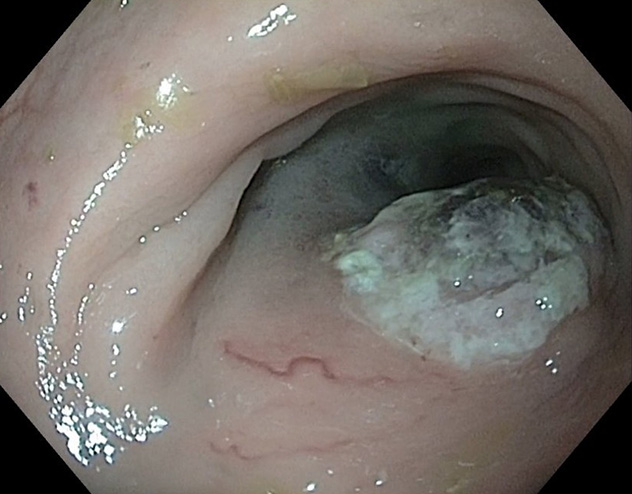
This image shows a preclinical colorectal cancer model.
Irreversible electroporation (IRE), also known as pulsed field ablation, is a highly selective, nonthermal modality for tissue ablation leading to irreversible cell death. Our innovative work involves identifying emerging endoscopic therapies for luminal cancers and creating preclinical cancer models.
In this research, a novel IRE device that uses a vacuum to draw target tissue into contact with the electrodes was mounted onto a colonoscope. The device was placed directly onto the cancer tissue to deliver therapy.
The colorectal cancer model used was a genetically modified pig with inducible transgenes ending for Kirsten rat sarcoma viral oncogene and tumor protein p53 driver mutation. Advancing this type of cutting-edge technology provides alternative options to manage cancers and precancerous diseases in patients.
Optical coherence tomography in Barrett's esophagus
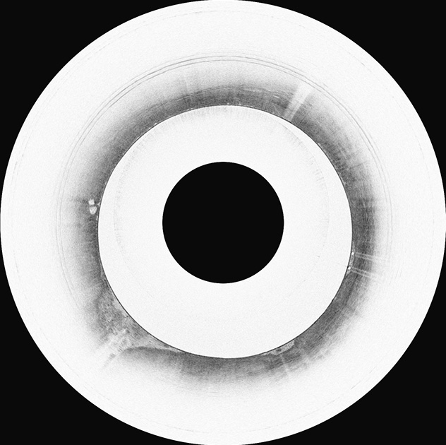
This is a representative image of Barrett's esophagus detected using optical coherence tomography.
Optical coherence tomography is an advanced imaging modality that provides high-resolution cross-sectional images of the human esophagus. This kind of tomography is used to screen for Barrett's esophagus while simultaneously detecting tissue microarchitectural changes associated with early esophageal neoplasia.
Integration of artificial intelligence enables semiautomated, in vivo interpretation of optical coherence tomography imaging data. Our work has focused on codevelopment and validation of optical coherence tomography systems for Barrett's esophagus screening and surveillance.
Key references
Other research projects
In addition to these projects, our lab has several other ongoing projects in partnership with medical device companies. These projects include the study of innovative approaches for noninvasive detection of colorectal polyps and cancer, weight-loss devices, and endoscopic closure devices to manage defects or perforation of the gut wall.