Microfluidic cell or explant cultures
Microfluidic co-culture systems
Microfluidic devices may be used to culture distinct cell types in close proximity to study how cellular interactions contribute to health or disease. Our team has developed and reported on a series of devices that may be used to study paracrine or juxtacrine interactions.
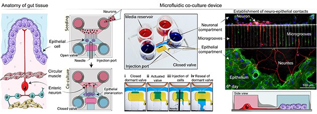
The figure below describes a device developed by our team where gut epithelial cells may be cocultured with enteric neurons.
 Studying neuro-epithelial interactions
The Cellular Microsystems and Biosensors Lab has developed a microfluidic coculture device to study how neuro-epithelial interactions contribute to gastrointestinal dysfunction.
This device may be used to investigate how neuro-epithelial connections impact gut function. From a technology standpoint, this microfluidic device addresses a problem of efficiently seeding gut organoids derived from patients by implementing an injection port to load cells in the active regions of the device.
Other devices our team is developing focus on investigating immune-epithelial cell interactions.
Microfluidic cell or explant cultures
There is a pressing need to move away from one-size-fits-all treatments and toward patient-specific therapy selection. Our team is interested in employing microfluidic devices to culture cells or tissue derived from patients for personalized medicine applications.
While multiple clinical areas stand to benefit from this approach, oncology is one field where the need is particularly pressing and compelling. Therefore, we have been using microfluidic devices for applications in cancer cell and explant cultures.
We find such devices useful for several reasons:
- They require less tissue.
- They are particularly well suited for clinical workflows involving cancer biopsies.
- Cells or tissue often survive better in microfluidic devices because of enhanced autocrine or paracrine signaling.
One type of microfluidic cancer culture device that the lab frequently uses contains microwells. This device helps organize cells into spheroids or organoids. This system has been used to successfully culture pancreatic, ovarian and liver cancer cells.
The figure below shows ovarian cancer cultures.
 Testing chemotherapies
The image on the left shows a microfluidic device integrating multiple cell culture chambers. This device allows for parallel testing of multiple chemotherapy types or concentrations. The image in the center shows cancer spheroids inside a microfluidic chamber. Each spheroid resides in its own microwell and may be monitored over time by microscopy. The image on the right shows immunofluorescence staining of spheroids for markers of cancer.
In addition to testing chemotherapies, this type of device has been used to assess cell-based immunotherapy.
Key publications
Choi D, Gonzalez-Suarez AM, Dumbrava MG, Medlyn M, de Hoyos-Vega JM, Cichocki F, Miller JS, Ding L, Zhu M, Stybayeva G, Gaspar-Maia A, Billadeau DD, Ma WW, Revzin A. Microfluidic organoid cultures derived from pancreatic cancer biopsies for personalized testing of chemotherapy and immunotherapy. Advanced Science. 2024.
De Hoyos-Vega JM, Yu X, Gonzalez-Suarez AM, Chen S, Mercado-Perez A, Krueger E, Hernandez J, Fedyshyn Y, Druliner BR, Linden DR, Beyder A, Revzin A. Modeling gut neuro-epithelial connections in a novel microfluidic device. Microsystems & Nanoengineering. 2023.
Dadgar N, Gonzalez-Suarez AM, Fattahi P, Hou X, Weroha JS, Gaspar-Maia A, Stybayeva G, Revzin A. A microfluidic platform for cultivating ovarian cancer spheroids and testing their responses to chemotherapies. Microsystems & Nanoengineering. 2020.
Zhou Q, Patel D, Kwa T, Haque A, Matharu Z, Stybayeva G, Gao Y, Diehl AM, Revzin A. Liver injury-on-a-chip: Microfluidic co-cultures with integrated biosensors for monitoring liver cell signaling during injury. Lab on a Chip. 2015.