Noninvasive optical imaging
The Optical Imaging Lab provides noninvasive imaging (fluorescent or bioluminescent) of living mice using the PerkinElmer IVIS Spectrum system. However, the autofluorescence in the gut, skin and fur of mice results in high background signals and can make imaging in living animals challenging. Hence, optical imaging is best performed with the bioluminescent modality.
Visit the Optical Imaging Lab's internal website (must be logged in to the Mayo Clinic network).
Bioluminescent imaging uses luciferase reporter genes to noninvasively image cells and viruses in living animals, enabling longitudinal studies. Bioluminescent imaging is highly sensitive due to the lack of background signals in mice.
General approach to imaging:
- Cells are transduced with lentiviral vectors, encoding luciferase prior to transplantation in animals.
- Confirm luciferase activity in transduced cells before using in animals, as shown.
- Implant cell lines into the animals.
- Remove the fur prior to imaging to enhance quality.
- Administer a dose of D-luciferin substrate to the mouse 10 to 15 minutes prior to imaging.
Investigators on Mayo Clinic's Rochester campus are welcome to use the PerkinElmer IVIS Spectrum system. For training and scheduling, contact:
Noninvasive SPECT, PET and CT imaging of mice and rats
Bioluminescent imaging, while highly sensitive, has poor resolution and works well only in mice due to attenuation of light photons by tissues. For higher resolution imaging in mice and rats, the Nuclear Medicine Molecular Imaging Shared Resource is available.
The Nuclear Medicine Molecular Imaging Shared Resource has a Siemens Inveon MicroPET/CT Scanner and an MILabs U-SPECT-II MicroSPECT/CT for high-resolution imaging of mice and rats. For example, numerous investigators at Mayo Clinic have been using the sodium iodide symporter (NIS) as a reporter gene to monitor gene transfer and tissue regeneration in living animals over time using these machines.
For more information, visit the Nuclear Medicine Molecular Imaging Shared Resource intranet site (must be logged in to the Mayo Clinic network).
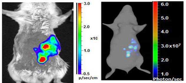 Bioluminescent image of a tumor
Bioluminescent imaging of an intraperitoneally implanted tumor cell line previously transduced with lentiviral vector encoding firefly luciferase (left) and the 3D rendering of the bioluminescent data (right). Source: Imanis-Life Sciences. Used with permission.
Related publications
Shen W, Patnaik MM, Ruiz A, Russell SJ, Peng K. Immunovirotherapy with vesicular stomatitis virus and PD-L1 blockade enhances therapeutic outcome in murine acute myeloid leukemia. Blood. 2016; doi:10.1182/blood-2015-06-652503.
Hickey RD, Mao S, Amoit B, Suksanpaisan L, Miller A, Nace R, Glorioso J, O'Connor MK, Peng K, Ideda Y, Russell SJ, Nyberg SL. Noninvasive 3-dimensional imaging of liver regeneration in a mouse model of hereditary tyrosinemia type 1 using the sodium iodide symporter gene. Liver Transplantation. 2015; doi:10.1002/lt.24057.
Moulay G, Ohtani T, Ogut O, Guenzel A, Behfar A, Zakeri R, Haines P, Storlie J, Bowen L, Pham L, Kaye D, Sandhu G, O'Connor M, Russell S, Redfield M. Cardiac AAV9 gene delivery strategies in adult canines: Assessment by long-term serial SPECT imaging of sodium iodide symporter expression. Molecular Therapy. 2015; doi:10.1038/mt.2015.78.
Miller A, Suksanpaisan L, Naik S, Nace R, Federspiel M, Peng K, Russell SJ. Reporter gene imaging identifies intraturmoral infection voids as a critical barrier to systemic oncolytic virus efficacy. Molecular Therapy — Oncolytics. 2014; doi:10.1038/mto.2014.5.