Facilities and equipment
The X-Ray Imaging Core is located on Mayo Clinic's campus in Rochester, Minnesota.
Whole-body CT scanning
The CT scanning service of Mayo Clinic's X-Ray Imaging Core is on Mayo Clinic's campus in Rochester, Minnesota. The imaging system is a commercial, whole-body Somatom Definition Flash dual-source CT scanner manufactured by Siemens Healthineers USA.
It is a leading technology CT system that can perform:
- 75-ms temporal resolution ECG-gated data acquisition.
- 0.4 mm isotropic voxel spatial resolution.
- Dual-energy CT.
- Reconstructions using image widths from 0.5 mm to 10 mm.
- Sequential, spiral and stationary table data acquisitions.
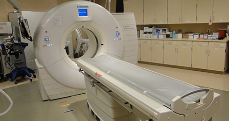
SOMATOM Definition Flash manufactured by Siemens Healthineers USA
Micro-CT scanning
The micro-CT scanning service of the X-Ray Imaging Core is on Mayo Clinic's campus in Rochester, Minnesota. The X-Ray Imaging Core has access to four micro-CT scanner systems that provide imaging services appropriate to the needs of various research applications. These resources provide high-resolution, nondestructive 3D mapping of a sample's X-ray attenuation properties.
- MCT4 scanner. This commercial scanner offers flexible geometric configurations and can support a wide range of custom applications, such as high-energy micro-CT and scatter experiments. The scanner is outfitted with a micro-focus X-ray source that has a tungsten anode. It provides three focus modes: focal spot size of 5 µm, 20 µm and 50 µm. It also provides a 40 kV to 150 kV range of operational voltages. The X-ray detector is a complementary metal-oxide semiconductor (CMOS) flat panel detector that offers a 15 cm-by-12 cm active area, 95-µm pixel pitch, three-pixel binning modes and a frame rate of up to 30 frames per second. Flexible positioning of the system hardware enables high-magnification applications yielding effective pixel sizes down to 5 µm.
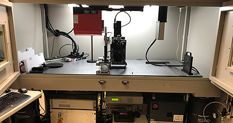
MCT4 specimen imaging scanner
- MCT6 scanner. This micro-CT scanner offers phase-contrast imaging in addition to traditional X-ray absorption imaging. A single acquisition can yield three contrasts: absorption, phase and dark field. Absorption is sensitive to high-density materials such as bone. Phase is sensitive to low-density materials such as soft tissue. And dark field is sensitive to scatter caused by small structures and edges. Incorporating phase-contrast imaging capabilities enhances preclinical imaging applications by allowing for the extraction of complementary X-ray attenuation and phase information that derive from unique interactions in various tissues. The scanner is outfitted with a micro-focus X-ray source that provides three focus modes: focal spot size of 5 µm, 20 µm and 50 µm. The optimal voltage for the phase-contrast grating interferometers is 40 kVp. The system has a 4-megapixel CMOS X-ray detector that has a cesium iodide scintillator and a pixel pitch of 23 µm.
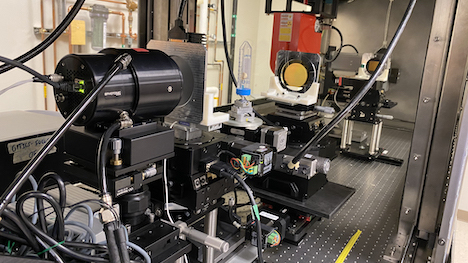
MCT6 phase-contrast micro-CT
- Bruker SkyScan 1272. This commercial scanner offers a compact system design that enables high-resolution micro-CT imaging of in vitro and ex vivo specimens. The scanner is outfitted with a micro-focus X-ray source that provides a 20 kV to 100 kV range of operational voltages using a tungsten anode with an automatic filter changer. The X-ray detector is an 11-megapixel charge-coupled device (CCD) detector with a fiber-optic-coupled scintillator and includes four pixel binning modes. The system has adjustable magnification settings and can yield effective pixel sizes from 6.35 µm down to 0.45 µm with the 1-pixel binning mode. The system can accommodate maximum specimen sizes of 75 mm in diameter and 70 mm in length. The scanner console includes installed vendor software that enables reconstruction, 2D-3D numerical analysis and 3D visualization.
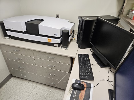
Bruker SkyScan 1272 scanner
- Bruker SkyScan 1276. This commercial scanner offers a compact system design with a rotating gantry to enable micro-CT imaging with high spatial and temporal resolution. These features accommodate a range of research applications, including ex vivo, in vitro and in vivo imaging scenarios. The scanner is outfitted with a micro-focus X-ray source that provides a 20 kV to 100 kV range of operational voltages using a tungsten anode with an automatic filter changer. The X-ray detector is an 11-megapixel CCD detector with a fiber-optic-coupled scintillator and includes four pixel binning modes. The system has adjustable magnification settings and can yield effective pixel sizes from 10 µm down to 3 µm with the 1-pixel binning mode. The system can accommodate maximum specimen sizes of 80 mm in diameter and 300 mm in length. The scanner console includes installed vendor software that enables reconstruction, 2D-3D numerical analysis and 3D visualization. Also, the scanner has an integrated physiological monitoring software for in vivo applications. An isoflurane anesthesia system is near the scanner for research procedure preparation.
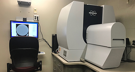
Bruker SkyScan 1276 scanner
Fluoroscopy
The mobile C-arm fluoroscopy service provided by the X-Ray Imaging Core on Mayo Clinic's campus in Rochester, Minnesota. The digital mobile Siemens Arcadis Avantic C-arm fluoroscope is used for portable fluoroscopy. The C-arm supports imaging needs for in vivo research procedures occurring in the nearby preparation lab, surgery suite and CT room.
The biplane fluoroscopy service of the X-Ray Imaging Core is on Mayo Clinic's campus in Rochester, Minnesota. Two Siemens Artis zee biplane fluoroscopes are available for use, and they are located in adjacent rooms. These imaging systems are routinely used to support in vivo research procedures involving interventional use and fluoroscopic guidance. Common examples include catheter insertion and balloon dilation, as well as injection of contrast, drugs or stem cells.
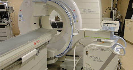
C-arm fluoroscope