Distal Radioulnar Joint
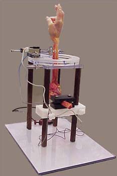
Figure 7: Test set-up for measuring DRUJ stability
Principal Investigator: Richard A. Berger, M.D., Ph.D.
Project Coordinator: Lawrence Berglund — berglund.lawrence@mayo.edu
Interosseous Membrane Contribution to Distal Radioulnar Stability
The interosseous membrane (IOM) is a contributing stabilizer of the distal radioulnar joint (DRUJ), but less so than the triangular fibrocartilage complex (TFCC). Previous studies have concentrated on the role of the IOM as a longitudinal stabilizer of the forearm. Although IOM injuries occur, they are difficult to diagnose. Results from previous studies have shown that disruption of the TFCC must occur in order for the DRUJ to dislocate, but studies have not clarified the IOM's role in restraining the DRUJ. It remains difficult to assess the status of the IOM clinically. We evaluated the role of isolated regions of the IOM related to dislocatability of the DRUJ in a cadaveric study (Figure 7).
From these studies, we conclude that palmar dislocation of the radius relative to the ulna implies a completely compromised TFCC but does not require an IOM injury, while a dorsal dislocation of the radius implies both a TFCC and distal IOM injury. Disengagement of the DRUJ implies an extended IOM injury including both distal and middle regions.
Role of Joint Capsule in DRUJ Stability
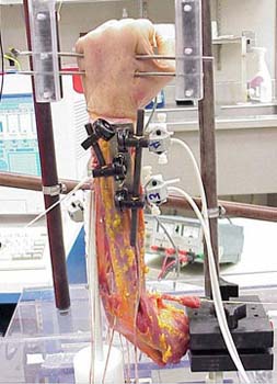
Figure 8: Kinematic study of DRUJ
This study was designed to specifically determine the contribution of the distal radioulnar joint (DRUJ) capsule as a stabilizer of the distal radioulnar joint. We analyzed the contribution of the joint capsule to DRUJ stability in a biomechanical study using human cadaveric upper extremities. The results demonstrated the effect of capsular injury on DRUJ joint stability, and the re-stabilizing effect of capsule shortening. The importance of the capsule to DRUJ stability should be considered when planning surgical procedures to restore the unstable DRUJ.
Kinematic Effects of Dorsally Angulated Distal Radius Fractures on the DRUJ
Dorsal angulation of the distal radius after healing of radial fractures is known to affect the constraints of the distal radioulnar joint (DRUJ). The purpose of this study was to examine the forearm torque required to achieve a full range of pronation and supination of the wrist for normal to increasing angles of radial dorsal angulation in a cadaveric model (Figure 8). Results would present criteria for performing corrective osteotomy.
In-Vivo Analysis of DRUJ Stability
A novel protocol for the radiographic assessment of stability of the distal radioulnar joint (DRUJ) was developed and introduced into clinical use in January, 2001. This protocol is centered on a device, which allows simultaneous bilateral computerized tomography imaging of the DRUJs. Imaging is done under various forearm positions both at rest and with active forearm resistance. The proposal calls for the recruitment of normal adult volunteers who will undergo torque strength testing and CT imaging of bilateral DRUJ's following the clinical protocol to assess the normal degree of DRUJ disassociation.