Our Research
The Neural Regenerative Research Lab studies ways to enhance peripheral motor nerve regeneration of autograft nerve, decellularized allograft nerve and biosynthetic nerve graft substitutes. To achieve these goals, our research uses cellular-based therapies (such as autologous and allogenic stem cells and exosomes) and immunomodulation of the local environment (such as surgical angiogenesis and local immunosuppression).
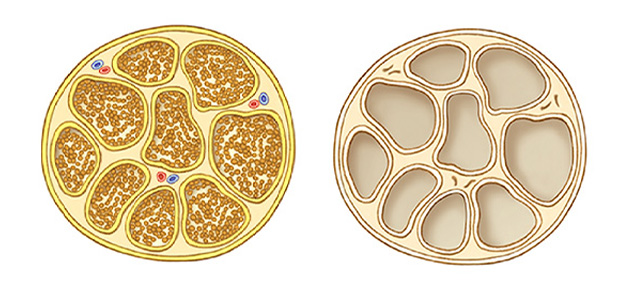 Nerve decellularization
An unprocessed nerve autograft is shown including all cellular structures and vascularization (left). After a five-day decellularization process, the nerve autograft is processed into a nerve allograft and stripped of all the cellular structures (right).
Improving patient care
Our basic science studies of nerve regeneration are born from our clinical experiences as surgeons performing brachial plexus injury repair. Despite enthusiasm for distal nerve transfers, the gold standard for most peripheral nerve injuries remains excision of the damaged portion and bridging with an interposition, cabled autologous nerve graft. Expendable sources of autograft are limited both in diameter and length, and harvesting results in permanent sensory deficits. Insufficient graft material and donor site morbidity limit the ability to optimally reconstruct nerve gaps, frequently compromising clinical outcomes, especially in brachial plexus injuries.
Our surgical practice at Mayo Clinic has benefited from advances in nerve basic science research and has provided fertile ground for clinical and translational research. Our multidisciplinary peripheral nerve care team has provided reconstructive treatment for thousands of patients. But the application of synthetic nerve conduits and commercially available decellularized nerve allografts have failed to overcome motor and mixed major motor and sensory nerve defects greater than 1.18 inches (3 centimeters) in length. Having an unlimited supply of patient-individualized nerve substitute remains a much-needed resource for our surgical practice to meet patient demand.
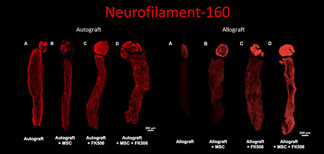 Nerve regeneration after treatment
Neurofilament-160 is a specific staining that shows the regeneration of neurites (in red) from the cell body (dorsal root ganglion; the circular top) into the nerve graft (rectangular attached to it). This is shown for nerve autografts and nerve allografts for similar treatments.
Developing an improved decellularized nerve allograft
To this end, our Neural Regenerative Research Lab has developed novel methods to decellularize nerves and enhance their regenerative potential by seeding them with adipose-derived mesenchymal stem cells and determining the role of surgical angiogenesis in peripheral nerve regeneration.
Studies in Dr. Shin's lab are designed to improve the effectiveness of nerve graft substitutes (such as acellular nerve allografts and biosynthetic nerve graft substitutes) by adding cellular components (stem cells and exosomes) and altering the local environment (surgical angiogenesis and local immunosuppression).
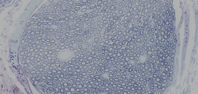 Histology of a nerve
Staining used to image the amount of regenerated nerves and the myelin sheath around the nerves as a measure of regeneration.
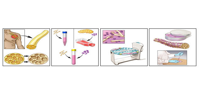 Stem cell-seeded nerve
Nerves are harvested, decellularized into acellular nerve allografts and seeded with cultured stem cells, after which a stem cell-seeded nerve is obtained. This nerve can be used for in vitro research or for nerve reconstruction surgery.