-
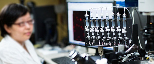
High-speed microscopy advances research
The lab uses high-speed fluorescent microscopy to determine responses of single cells and tissues to mechanical and chemical stimuli.
-
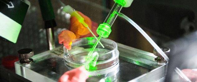
What do ion channels do in the gastrointestinal tract?
The lab uses single-molecule, whole-cell conventional electrophysiology and optogenetics to better understand gastrointestinal physiology.
-
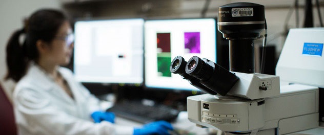
Seeing is believing
Confocal and super-resolution microscopy are valuable tools used in the lab’s studies.
-
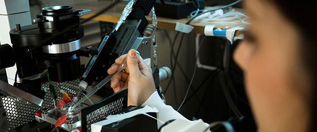
Novel techniques to study gastrointestinal physiology
The lab develops novel techniques for studying mechanosensitive ion channels in the gastrointestinal tract.
Overview
The Gastrointestinal Mechanotransduction Laboratory led by Arthur Beyder, M.D., Ph.D., studies the molecular mechanisms of gastrointestinal function in health and disease. The lab seeks to provide novel diagnostic and therapeutic tools for patients with functional gastrointestinal diseases.
Functional gastrointestinal diseases, such as irritable bowel syndrome (IBS) and functional dyspepsia, are common but difficult to diagnose and treat because the mechanisms are poorly understood. The lack of high-quality diagnostic tools and targeted treatments is frustrating for both the patient and physician.
Dr. Beyder and his colleagues focus on the molecular mechanisms of gastrointestinal motility. It is well-known that mechanical forces affect gastrointestinal function and mechanical sensitivity is abnormal in many patients with functional gastrointestinal diseases. Therefore, the work in Dr. Beyder's lab specifically focuses on understanding how mechanical forces impact cellular function both in health and disease. Dr. Beyder's lab studies mechanosensitive ion channels, a type of mechanical sensor in cells. Mechanosensitive ion channels are critical for normal gastrointestinal function.
The lab uses a wide variety of techniques. Studies start at the cellular and molecular level, using electrophysiology as the foundation, and complementary techniques ranging from immunohistochemistry to single-cell genomics. Conventional microelectrode-based electrophysiology and optogenetics are used as well. Tissue function profiling is accomplished using Ussing chambers for secretion and muscle bath for contractions.
Finally, to understand how stimulation at the molecular level impacts animals, a variety of transgenic mouse models are used to do live studies. Mechanical stimulation is integrated into these experiments with a variety of approaches, such as pressure clamp and flexible substrates.