Research
Our research team in the CT Clinical Innovation Center works to develop and evaluate new CT imaging technology and clinical applications, with a focus on quantitative applications of computerized tomography.
CT technology
This image shows our current CT scanner, which is a Siemens NAEOTOM Alpha.
Innovation is driven by new ideas, new opportunities and new technology. We evaluate and bring into clinical use the latest CT scanner technology available from our industry collaboration partner, Siemens Healthineers.
Since 2004, we have installed, tested and integrated into our clinical practice several CT systems that were the first in the United States:
- Sensation 64.
- Definition Dual Source.
- Definition AS+.
- Definition Flash.
- SOMATOM Force.
- SOMATOM CounT.
- SOMATOM Count Plus.
- NAEOTOM Alpha.
Deep learning noise reduction
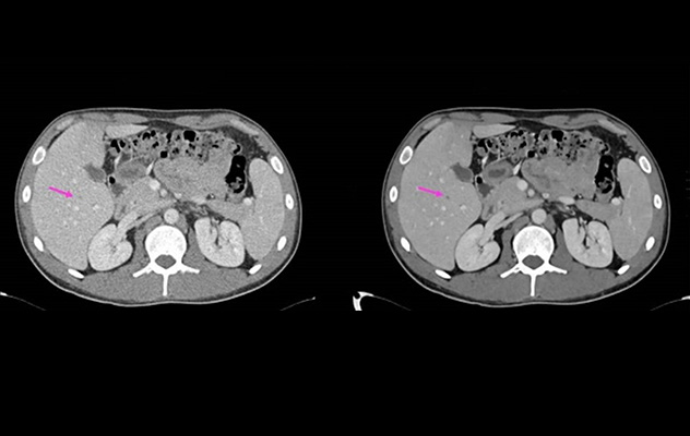
This image compares two CT scans of the liver. The scan on the left is more grainy (noisy) and hard to read. The scan on the right has been improved using our artificial intelligence-powered tool, making it much clearer. A small dark spot in the liver, marked by the red arrow, is easier to see in the improved image.
Our deep learning technology improves the quality of scans. Our artificial intelligence (AI)-powered tool helps make grainy (noisy) images look more clear, making them easier to read and interpret.
Super resolution
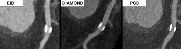
This image compares CT scan quality. The scan on the left shows a heart artery with calcium buildup, but it looks blurry and bigger than it really is. The image on the right is much clearer because it was taken with a more advanced scanner. The image in the middle, processed with DIAMOND, more accurately shows the calcium buildup, just like the high-tech scan on the right.
Our DIAMOND technology uses artificial intelligence to make regular CT scan images clearer and sharper.
Newer, more-advanced scanners can render much clearer images, but these better CT machines aren't widely available yet.
Our DIAMOND technology helps overcome that challenge by taking a scan from a regular CT and improving it so the images are much more clear.
Dose reduction
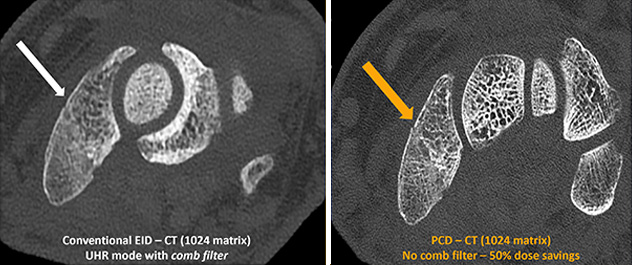
Two scans of a fractured wrist, indicated by an arrow, show differences in CT quality. The version on the right was taken with a new type of CT scanner called photon-counting detector CT, which renders crisper images with only half the radiation dose.
Despite the tremendous medical benefits of CT imaging, concerns remain about the potential for negative long-term effects of radiation exposure.
To address these concerns, our team develops and tests dose-reduction technologies to limit radiation exposure.
Our studies include:
- Image-space denoising.
- Projection-space denoising.
- Iterative reconstruction.
- Deep learning denoising.
- Spectral optimization.
- Automatic exposure control systems.
- Novel detector technology.
Multienergy CT
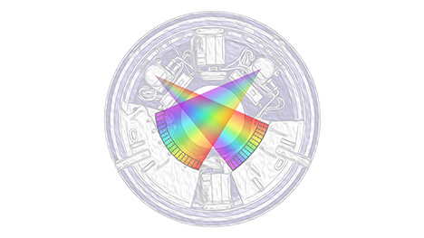
Our current photon-counting detector CT scanner uses two X-ray beams, each of which brings color information into the typically greyscale world of CT images. The X-ray beams in this artistic rendering of a CT machine are colored in the shades of a rainbow to show that they bring new information to CT scanning, just as color TV did for black-and-white TV.
Conventional CT imaging sees the body in shades of grey. Two materials of different composition can have the exact same brightness if their densities compensate for the differences in atomic number. A classic example is the difficulty in differentiating bone and calcium deposits from iodinated blood vessels.
To overcome this limitation of CT and open up new clinical applications focused on determining material composition, a second set of attenuation measurements is required. This second measurement is taken using a different X-ray energy spectrum. Commercial CT systems with this capability are known as dual-energy CT systems.
Multienergy CT can also be accomplished by differentiating photon energies at the detector using photon-counting energy-resolving detectors, such as CdTe and CZT. Multienergy CT brings color information to what are otherwise black-and-white images.